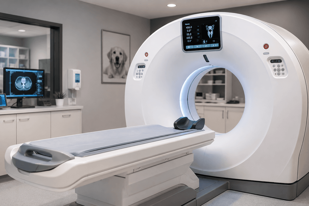
In the world of veterinary diagnostics, the journey often begins with a physical exam, followed by bloodwork and traditional imaging like radiographs (X-rays) or ultrasound. These tools are invaluable for a "first look" at your pet's internal health. However, as medicine advances, veterinarians are increasingly turning to Computed Tomography (CT) to solve complex medical mysteries.
If your veterinarian has recommended a CT scan after initial imaging, it is because they need a higher level of detail to provide the most accurate diagnosis and treatment plan. While X-rays provide a 2D "snapshot" and ultrasound offers a dynamic close-up view, CT provides a high-resolution, 3D anatomical map (Ohlerth & Scharf, 2007).
Computed Tomography, often called a "CAT scan," uses a rotating X-ray beam and advanced computer processing to create "slices" of the body. Unlike a traditional X-ray, which flattens the body into a single plane, causing organs to overlap in a phenomenon called "summation" (Meomartino et al., 2021), CT allows a specialist to look through the body layer by layer (Greco et al., 2022).
While ultrasound is excellent for looking inside soft tissue organs (Won et al., 2015), it can be limited by gas or bone interference. Conversely, while X-rays are the gold standard for bone, they lack the sensitivity to detect subtle soft-tissue changes (Harding et al., 2025). CT fills these gaps, particularly in the following clinical scenarios:
- Complex oncology (cancer) staging: CT is often the preferred choice for "whole-body" staging to determine if a tumor has spread (metastasized) to the lungs or distant lymph nodes (DE Almeida et al., 2025). It is significantly more sensitive than chest X-rays for finding small lung nodules (Forrest, 2016).
- Acute polytrauma: In cases of severe trauma, such as a "hit by car" incident, whole-body CT allows for a rapid, comprehensive assessment of internal bleeding, rib fractures, and spinal injuries that might be missed on standard X-rays (Dozeman et al., 2020). While this is nice to have, access may be limited in the emergency setting.
- Vascular and heart anomalies: Using CT angiography, the injection of a contrast dye, specialists can visualize blood flow to identify congenital defects like liver shunts (portosystemic shunts) or life-threatening blood clots in the lungs (pulmonary thromboembolism), among other abnormalities (Jackson et al., 2024; Goggs et al., 2014).
- Neurological and spinal issues: While MRI is often best for the brain and spinal cord, CT can also be effective for identifying acute spinal cord compression, "slipped" discs, or infections of the vertebrae, or discospondylitis (Dennison et al., 2010; Gomes et al., 2022).
- Critical GI obstructions: While ultrasound is excellent for many foreign bodies (Tyrrell & Beck, 2006), CT can be utilized to definitively diagnose mechanical obstructions when traditional imaging results remain inconclusive (Miniter et al., 2019).
In addition to soft tissue and vascular evaluation, CT is the definitive gold standard for assessing complex orthopedic injuries. While X-rays provide a preliminary view of bone health, they often fail to capture the full extent of "comminuted," or multi-fragment, fractures, particularly in anatomically dense regions.
- Pelvic and spinal fractures: CT allows for 3D reconstruction of the pelvis, which is essential for surgical planning to ensure proper alignment of the weight-bearing axis (Dozeman et al., 2020).
- Jaw and facial trauma: For fractures of the mandible or maxilla, CT provides unparalleled detail of the dental roots and temporomandibular joints, which are frequently obscured by the "summation" of the skull on standard radiographs (Chai et al., 2017). This can allow for more precise prognostic information and surgical fixation or stabilization options.
- Long bone and joint involvement: Complex fractures may require high-detail anatomic information to optimize reconstruction by a surgeon. When a fracture extends into a joint (intra-articular), CT may be necessary to map the joint surface, helping surgeons minimize the risk of secondary osteoarthritis (Meomartino et al., 2021).
One of the most powerful applications of CT is its use in guiding minimally invasive procedures. Much like ultrasound-guided sampling (Holter et al., 2023), CT allows a specialist to perform highly precise "CT-guided" biopsies of lesions in difficult-to-reach areas, such as the nasal cavity or deep within the pelvic canal (Laborda-Vidal et al., 2022). This provides a path to diagnosis that previously relied solely upon major surgery (Vignoli & Saunders, 2011).
Because a single CT scan can generate thousands of individual images, the acquisition and interpretation require specialized expertise. Access to CT scanners is typically reserved for specialty hospitals, though some more advanced and larger general practices may have these machines in-house. The resulting images are typically reviewed by a board-certified radiologist to ensure no subtle detail is missed (Ohlerth & Scharf, 2007).
Furthermore, most CT scans require either heavy sedation or general anesthesia to ensure the patient remains perfectly still during the brief scan. New protocols, such as "respiratory-gated" CT, are being developed to allow for better imaging in awake or lightly sedated patients with respiratory disease (Navarro et al., 2025).
The decision to move to CT is often driven by the need for surgical planning or to find an answer that remains "hidden" on X-rays and ultrasound (Shanaman et al., 2013). Accessing this level of care typically requires a referral to a specialty hospital or a diagnostic center equipped with advanced imaging capabilities.
If your primary care veterinarian has recommended an advanced workup, Vetmodo can help you locate specialty facilities and board-certified experts who offer CT and other advanced veterinary imaging services. Ensuring your pet has access to the right modality at the right time is the cornerstone of modern, evidence-based veterinary care. Search Vetmodo today to find a specialty center near you.