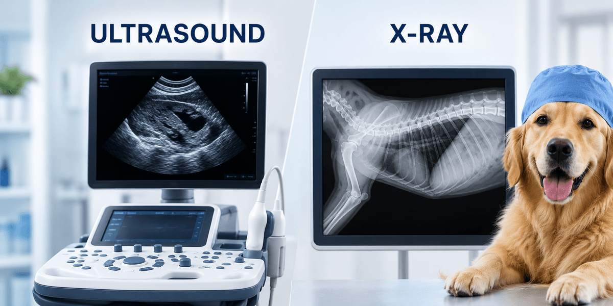
When a pet presents with clinical signs such as vomiting, lethargic behavior, or respiratory distress, medical imaging is the primary tool for definitive diagnosis. While radiographs (X-rays) and ultrasound are both forms of diagnostic imaging, they utilize different physical properties to visualize internal structures and are frequently used as complementary, rather than redundant, modalities (Meomartino et al., 2021).
X-rays, or radiographs, are often the initial step in a diagnostic workup due to its ability to capture a broad survey of the body. It excels at evaluating high-contrast structures such as bone and air-filled organs. In the context of the thorax, radiographs are the standard for assessing the distribution of lung patterns and heart size (Ward et al., 2018).
However, radiographs are limited by "summation," where overlapping soft tissue structures create a single silhouette. This makes it difficult to distinguish individual abdominal organs if fluid is present or if the organs are of similar density (Won et al., 2015). While X-rays are useful for identifying clear mechanical obstructions, such as a radio-opaque foreign body, they may be less accurate for "occult" obstructions compared to ultrasound (Sharma et al., 2011); (Tyrrell & Beck, 2006). Furthermore, serial radiographs alone do not always increase the accuracy of a diagnosis for certain gastrointestinal obstructions (Elser et al., 2020).
Ultrasound uses high-frequency sound waves to visualize the internal architecture of soft tissues, bypassing the summation issues of X-rays. This allows the clinician to evaluate the thickness of the gastrointestinal wall and the internal texture of organs like the liver and spleen (Shanaman et al., 2013).
In oncological cases, ultrasound is a critical tool for restaging. Research indicates that radiographs have limited cost-effectiveness when compared to the superior detail provided by ultrasound for identifying the spread of solid, soft-tissue tumors (Bevelock et al., 2025). Additionally, in cases of complex systemic disease like immune-mediated hemolytic anemia (IMHA), ultrasound is frequently utilized to rule out underlying triggers that radiographs might miss (Andres et al., 2019).
The most significant clinical advantage of ultrasound is the ability to perform real-time, guided sampling of lesions. When an X-ray identifies an abnormality, ultrasound can be used to visualize that specific area and guide a needle for a fine-needle aspirate (FNA) or biopsy.
This technique allows for the collection of cellular samples from the gastrointestinal wall, spleen, or other abdominal masses (Henry et al., 2024); (Holter et al., 2023). Because the clinician can see the needle's path on the monitor, they can avoid major blood vessels and adjacent organs, maintaining a high safety profile with minimal complications (Léveillé et al., 1993); (Wypij, 2011).
There is a fundamental difference in how these images are interpreted and acquired. Radiographs are standardized images that are frequently reviewed via teleradiology, where a board-certified specialist reviews the files remotely (Meomartino et al., 2021).
Conversely, ultrasound is a dynamic, operator-dependent examination. Acquisition requires advanced training to manipulate the probe and interpret the "live" feed. While basic image quality may be similar across various levels of expertise in healthy patients, the identification of subtle or complex pathology typically requires a high degree of sonographic skill (Lindquist et al., 2021); (Zelachowski et al., 2024).
Radiography vs. Ultrasound: Common Indications for Your Pet
- Radiography (X-rays):
- Skeletal evaluation for fractures or osteoarthritis (Harding et al., 2025).
- Survey of the chest for heart size and lung patterns (e.g. heart failure, pneumonia, other lung diseases) (Ward et al., 2018).
- Screening for radio-opaque gastrointestinal foreign bodies (stones, metal, bone).
- Screening for urinary bladder stones
- Evaluating gas patterns within the stomach and intestines (Sharma et al., 2011).
- Diagnostic Ultrasound:
- Detailed evaluation of organ architecture (liver, gallbladder, spleen, kidneys, pancreas, adrenals,urinary bladder, prostate/reproductive organs, lymph nodes).
- Measurement of gastrointestinal wall layering and thickness (Henry et al., 2024); identification detailed evaluation of intestinal foreign material/blockages.
- Guided sampling (FNA/biopsy) of internal masses (Holter et al., 2023).
- Identifying fluid (effusion) within the abdomen or chest.
*note: this is not a comprehensive list
The relationship between radiography and ultrasound is fundamentally symbiotic. While a radiograph provides the anatomical context and "big picture" measurements (Lamb & Nelson, 2015), ultrasound provides the internal detail and the clinical pathway for sampling. One identifies the location of the anomaly, while the other characterizes its nature.
However, accessing both modalities can be a logistical challenge for pet owners. While nearly all veterinary practices offer in-house radiography, diagnostic ultrasound often requires advanced equipment and a clinician with specific expertise in image acquisition. Finding a facility that offers both can be the difference between a quick answer and a multi-day referral process. Vetmodo helps bridge this gap by connecting pet owners with veterinary professionals who possess the advanced training and specialized equipment necessary to offer these critical, complementary diagnostic services in one location.